• MIT researchers have developed a novel way of creating three-dimensional, nanoscale versions of larger objects.
• They patterned it after an existing technique for enlarging samples, which their lab developed years ago.
• The new method, called implosion fabrication, has many potential applications in robotics, medicine, and optics.
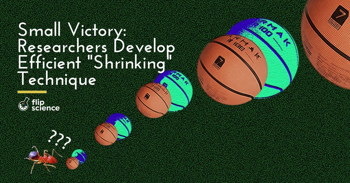
Ant-Man’s Pym particles present too much of a physics-defying problem to exist in real life, but that doesn’t preclude other out-of-the-box approaches to “shrinking” things. And that’s exactly what researchers at the Massachussets Institute of Technology (MIT) have developed: a novel way of creating three-dimensional, nanoscale versions of larger objects.
The new technique, called implosion fabrication, involves using a laser to create a pattern on a polymer scaffold. Afterwards, they attach other materials to the scaffold to replicate structures that are up to a thousand times bigger, “shrinking” them, in a sense.
From a giant leap to a small step
What makes this different from existing techniques for nanoscale printing? For starters, this new technique is faster and easier. Furthermore, it isn’t restricted to specialized materials (like polymers and plastics), as researchers can use metals, quantum dots, and even DNA for patterning. Moreover, existing techniques can only create self-supporting structures (like a solid cube, for instance, but not a hollow sphere or a chain link).
According to the recently published study, the authors adapted a technique called expansion microscopy, which they developed years ago. By enveloping tissue with a hydrogel and expanding it, they can produce high-resolution imaging using a microscope. They can also generate 3D visualizations of cells and tissues without expensive equipment.
The researchers realized that the technique could also work the other way around. Using an extremely absorbent material made from polyacrylate (the superabsorbent polymer found in commercial diapers), they created a scaffold. Afterwards, they bathed this in a solution with molecules of fluorescein, an organic compound used as a coloring agent. They then attached the molecules to specific points in the structure using laser light. The molecules serve as anchors for other molecule types, enabling the researchers to add them to the scaffold.
“It’s a bit like film photography — a latent image is formed by exposing a sensitive material in a gel to light,” says lead author Daniel Oran. “Then, you can develop that latent image into a real image by attaching another material, silver, afterwards.”
Shrinking the gap
At present, the researchers have been able to make objects about 1 cubic millimeter in size, with a 50-nanometer resolution. With further refinement of the process, however, better resolution will be possible.
Some of the practical applications of this new technology are in optics (for making specialized lenses for studying light or improving lenses in microscopes and camera phones) and robotics (for creating extremely small electronics or robots).
And the best part? It only requires equipment that most biology and materials labs already use.
“There are all kinds of things you can do with this,” according to senior author Edward Boyden. “Democratizing nanofabrication could open up frontiers we can’t yet imagine.”
Cover photo: Pexels
References
- http://news.mit.edu/2015/enlarged-brain-samples-easier-to-image-0115
- http://news.mit.edu/2018/shrink-any-object-nanoscale-1213
- http://science.sciencemag.org/content/362/6420/1281

Author: Mikael Angelo Francisco
Bitten by the science writing bug, Mikael has years of writing and editorial experience under his belt. As the editor-in-chief of FlipScience, Mikael has sworn to help make science more fun and interesting for geeky readers and casual audiences alike.